Demystifying Endoscopic Imaging: From Light Capture to Screen Display
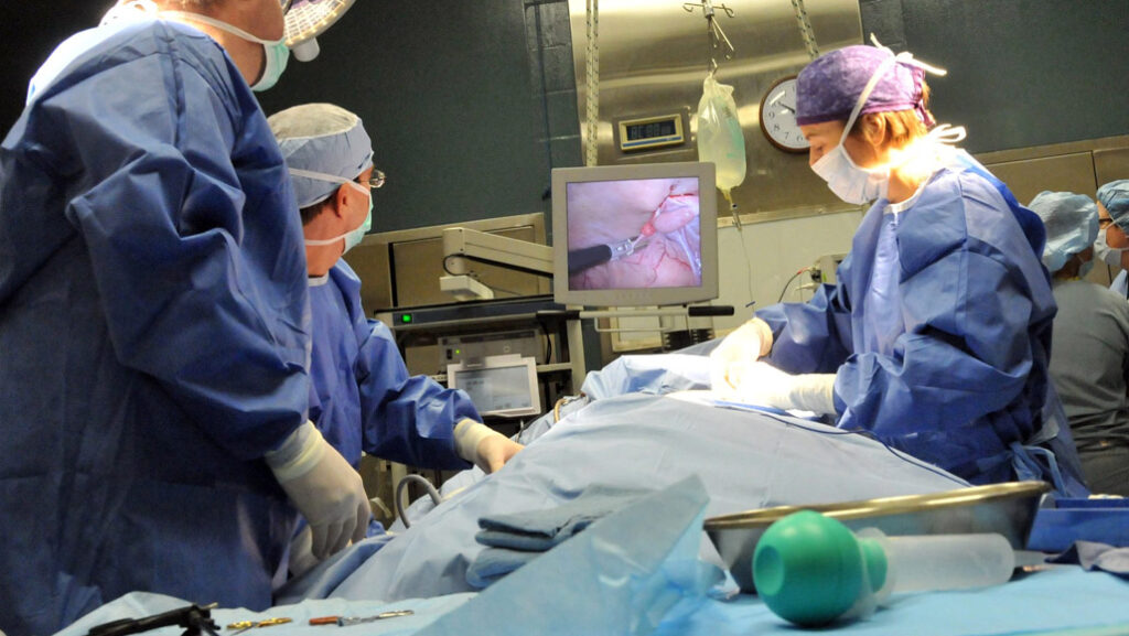
The ability to visualize the internal landscapes of the human body rests on the sophisticated synergy between optics, electronics, and illumination within an endoscope. Understanding these core systems reveals how clinicians achieve the clear, real-time imagery vital for accurate diagnosis and therapy.
The Fundamentals of Optical Image Capture
At the very tip of the endoscope lies the critical optical system responsible for initial image capture. Traditional rigid endoscopes employ a series of precisely aligned rod lenses to transmit the image along the instrument's length. In contrast, modern video endoscopes have largely adopted two advanced methods: high-density fiber optic bundles for image transmission or, more commonly, miniaturized CMOS sensors placed directly at the distal end to capture the image digitally.
The pursuit of exceptional clarity has led to technologies like 4K ultra-high definition imaging, which utilizes pixel arrays at a microscopic scale to deliver dramatically enhanced resolution. This front-end optical assembly, comprising the objective lens and subsequent lens groups, is meticulously engineered to minimize optical aberrations and color distortion. This precision ensures a uniformly sharp and accurate image from the center to the edges of the field of view.
The Journey of the Image: Transmission and Processing
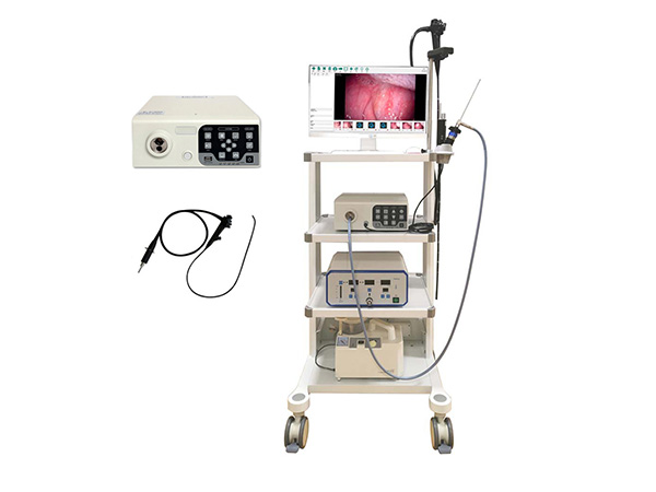
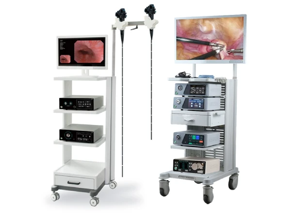
Once light from the internal cavity is focused, the image must be conveyed and refined. In a fiber optic-based system, the coherent bundle carries the optical image to a camera head. In a fully digital video endoscope, the CMOS sensor immediately converts the optical image into electrical signals.
These raw signals are then transmitted via internal wiring to a dedicated video processor. This processor performs a series of enhancements—adjusting color balance, improving contrast, reducing noise, and sharpening details—to optimize the image for clinical assessment. The processed video signal is finally output to a high-resolution medical monitor. The entire chain utilizes high-speed data interfaces to ensure the display is virtually instantaneous, providing true real-time feedback to the operator without perceptible delay.
Illumination: Shedding Light on the Procedure
A brilliant and reliable view depends entirely on the endoscope's illumination system. Modern endoscopes universally use "cold" light sources, primarily high-intensity LEDs or xenon lamps, which produce bright light while generating minimal heat at the source. This light is channeled from the generator through a dedicated bundle of light-carrying fibers running the length of the endoscope to its distal tip.
LED technology has become predominant due to its significant advantages: high luminous efficiency, low power consumption, exceptional longevity, and instant on/off capability. These systems feature adjustable intensity controls, allowing the surgeon to tailor brightness to the specific tissue and procedural needs. Furthermore, advanced optical coatings on lenses and windows within the system are applied to suppress internal reflections, glare, and "ghost" images, thereby elevating image contrast and overall visual clarity for more confident navigation and intervention.
In Conclusion: The Engine of Precision
The seamless video feed seen during an endoscopic procedure is the product of intricate and coordinated systems. From the advanced optics that first capture the image, through the rapid digital processing and transmission, to the powerful yet controllable illumination that makes it all visible, each component is engineered for precision and reliability. Continuous innovation in these core technologies directly translates to improved visualization, enhancing procedural accuracy and patient outcomes.